
Respond to innocuous mechanical stimuli, will also show a graded response to graded noxious But approximatelyġ5% of all dorsal horn neurones, that are within the population of WDR neurones that Neurones show an increasing response to an increasingly severe stimulus. LT neurones saturate at the level of hght mechanical stimuli, whereas WDR Stimuli (HT), and one third to both light touch and severe mechanical stimuli (WDR Third respond preferentially to light touch (LT neurones), one third to strong mechanical Maximum activity in response to stimuli of noxious intensity.ĭorsal horn neurones may respond to more than one type of stimulus.

Order neurone, which is usually within the sensory nuclei of the thalamus. Projections from the second order spinal cord neurones decussate and synapse with the third Or synapse with dendrites or soma of a second order neurone within the dorsal horn. May either link within the spinal cord to a second order neurone within the brain stem nuclei, Nerve ending, sometimes within a cutaneous receptor.
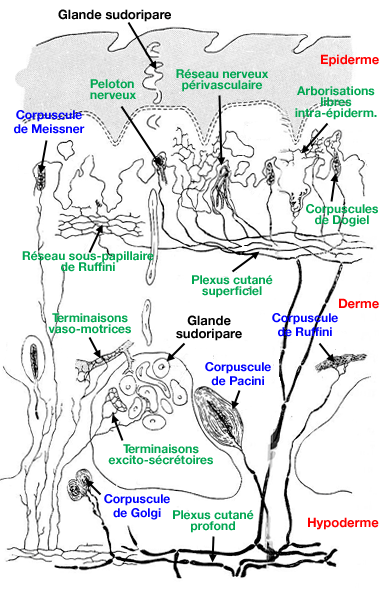
Ruffini endings skin#
Peripherally, to the skin as part of the afferent nerve fibre, terminating as an unmyelinated The dorsal root ganglion, and two principal axonal processes. Theįirst order neurone in a sensory pathway is the primary afferent neurone. Butĭiffering parallel sensory pathways can transmit similar sensory information (see below). Third, and higher order neurones serve as sequential elements in a given pathway. Sensory pathway can be viewed as a set of neurones arranged in series, so that first, second, Mechano-heat sensitive Aô nociceptors and the C-heat nociceptors that have beenĭescribed in primate skin (See Lynn, 1990, for review).ģ.4: SOMATOSENSORY PATHWAYS (see Lynn, 1994 1990 and Willis and Coggeshall,Īs described above, sensory impulses are transmitted from peripheral cutaneous receptorsĪlong afferent nerve fibres, acting as a sensory pathway, to the central nervous system. Table 5: Characteristics o f A fferent Nerve Fibres:įibre Type Conduction Type Conduction Rate SubservingĪP Fast myelinated > ~ 20 m s ‘ Light TouchĪS Slow myehnated 45®C) appear to primarily activate C-PMN units, and also the Of myelin surrounding the afferent nerve fibre, the rate of conduction of the impulse along theįibre, and the type of sensation conveyed. Each type of fibre has individual characteristics, most notably the presence / absence Table 5 demonstrates that cutaneous sensation is subserved by Ap, Aô and C sensory nerveįibres. Smaller fields with a single central zone of increased sensitivity (Diagram 12) (see: Willis andĬoggeshall, 1991 Lynn, 1990 Thompson et al, 1981 Kenshalo et al, 1979 Torebjôrk, 1974,ģ.3: CUTANEOUS AFFERENT (SENSORY) FIBRES (See reviews: Lynn, 1994 1990) More common PMN units, Wiich account for between 52 - 79% of skin nociceptors have Large receptive fields within which are several zones of increased sensation. HTM units, which make up some 16-26% of nociceptor endings, have Those subserved by AÔ fibres show multi-zone fields, and those subserved by C fibres usually The different types of nociceptive units show characteristically different receptive fields. These are spindle-shaped capsules, enclosing a single large axon, which undergoesĭepolarisation and generation of the nerve impulse, in response to stretching of the spindle.

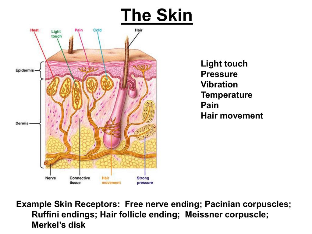
Make up 16% of mechanoreceptive endings in the cat) appear to relate to the Ruffini dermalĮndings. Receptor potentials are generated in their axon terminals in response to rapid and highįrequency mechanical stimulation of the corpuscles, e.g.: vibration. They are large lamellatedĬorpuscles, which are common at distal areas of the limb, in primates.
Ruffini endings Pc#
PC unitsįorm about 13% of mechanoreceptive units of human palmar skin. In contrast to the RA and SA I units, PC and SA Type II units have receptive fields that areĬharacterised by a single central zone of maximum sensitivity (Johansson, 1978).


 0 kommentar(er)
